Chest Imaging
Analysis of Misdiagnosis by CT and MRI of Solid Pseudopapillary Tumor of the Pancreas in the Elderly Patient
Author:ZHAN Qian, WANG Tie-gong, HUANG Ting.
affiliation:Department of Radiology, Shanghai Changhai Hospital, Shanghai 200433, China
PDFAbstract
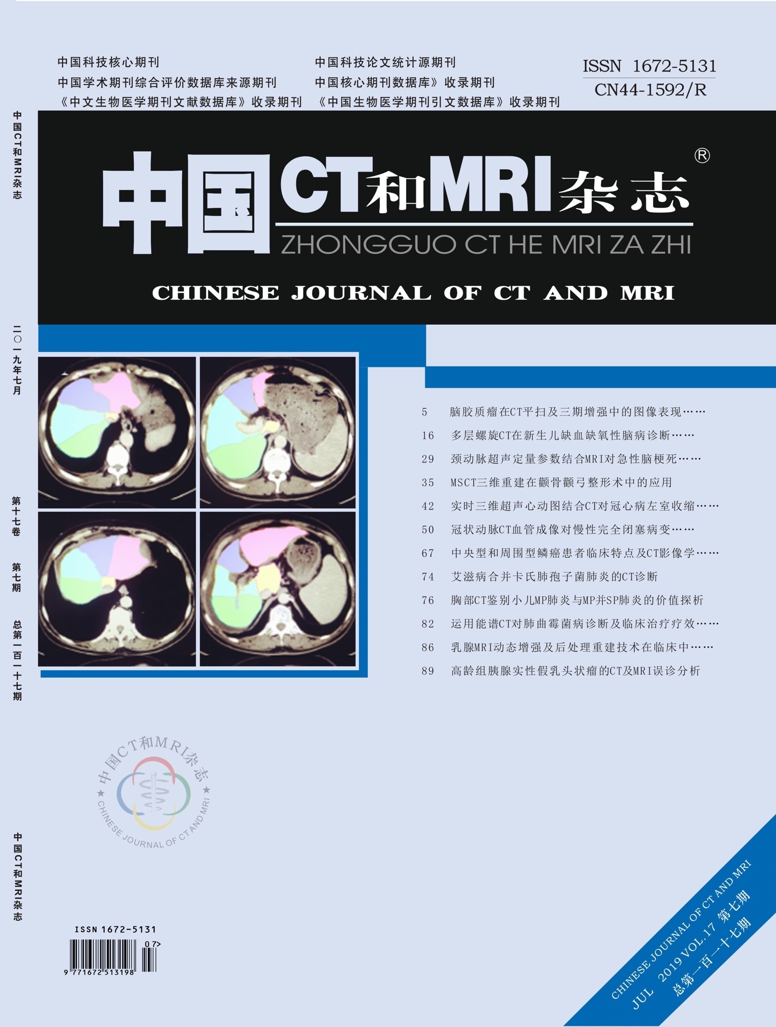
Objective To investigate CT and MRI features of solid pseudopapillary tumor of the pancreas in the elderly group. Methods Imaging findings in 9 elderly patients of solid pseudopapillary tumor of the pancreas confirmed by pathology and misdiagnosed by radiologic imaging (including CT and MRI). Results Group CT: A total of 5 cases. 2 cases were large (the longest diameter>10cm), internal structure was uneven (cystic necrosis, hemorrhage and calcification could be observed. 3 cases were small (the longest diameter less than 5cm), the internal structure was uniform. 5 cases of lesions showed less clear boundaries. After enhancing lesions compared with normal pancreatic tissue: (1) A low degree of enhancement. (2) Peak enhancement appeared later. Group MRI: A total of 4 cases. Showed a cystic lesion signal change, 3 cases of intracapsular lesions have separated, 1 case of cystic lesions more uniform composition. After enhancing solid components lesions progressive enhancement. Conclusion Preoperative diagnosis of solid pseudopapillary tumor of the pancreas in the elderly group could be marked by radiologic imaging.
【Keyword】Pancreas; Solid Pseudopapillary Tumor; Elderly Patient; CT; MRI
【Chart number】R445.2;R445.6
【Document Identification Number】A
【DOI】1 0 . 3 9 6 9 / j . i s s n . 1 6 7 2 - 5131.2019.07.027
Chinese journal of CT and MRI
th17Volume, th 7 Issue
2019Year07Month
Related articles
![]()
Editor email:(1)中国CT和MRI杂志:ctmri@vip.163.com;(2)罕少疾病杂志:hs1999@vip.163.com;(3)官网系统自动发论文录取通知书(注意!请勿回复或发送邮件到此邮箱):send@diagnoschina.com
Contact number:0755-83695203
Our address:

 English
 简体中文
简体中文 logged in
 Wechat
 TouTiao